- Have any questions?
- [email protected]
Brain Herniation: Types, Symptoms, Causes and Treatment
Brain herniation, sometimes referred to as hernia on the brain, is a life-threatening condition that occurs when brain tissue shifts or moves within the skull. This serious medical emergency can result from various causes, including severe head injuries, strokes, or brain tumours.
This article delves into the complexities of brain herniation, covering its types, causes, symptoms, and potential complications. We'll explore the diagnostic methods used to determine this condition & discuss the available treatment options. Additionally, we'll provide insights on when to seek medical attention and highlight preventive measures to reduce the risk of brain herniation.
What is Brain Herniation?
Brain herniation is a life-threatening condition that occurs when brain tissue shifts or moves abnormally within the skull. This serious medical emergency happens when increased intracranial pressure causes the brain to be pushed through openings in the rigid intracranial barriers. These barriers include the falx cerebri, tentorium cerebelli, and foramen magnum.
There are several types of brain herniation. The most common types include:
- Subfalcine Herniation: Brain tissue moves underneath the falx cerebri in the middle of the brain.
- Transtentorial Herniation: This type has two subtypes:
- Descending Transtentorial (Uncal): The uncus, part of the temporal lobe, shifts downward.
- Ascending Transtentorial: The cerebellum and brain stem move upward through the tentorium cerebelli.
- Tonsillar Herniation: The cerebellar tonsils move downward through the foramen magnum.
- Central Herniation: Both temporal lobes herniate through the tentorial notch.
Causes and Risk Factors of Brain Herniation
Brain herniation syndrome has multiple causes and risk factors that can lead to increased intracranial pressure. Some of the causes are:
- Presence of Space-occupying Lesions Within the Skull: These lesions can include haematomas, such as traumatic epidural and subdural haematomas, brain tumours and abscesses. These masses exert pressure on the surrounding brain tissue, potentially causing it to shift.
- Cerebral Oedema: It can result from various conditions. Malignant infarction, a severe form of ischaemic stroke, can cause substantial brain swelling.
- Infections: These include abscesses and empyemas, which can also lead to increased intracranial pressure and subsequent herniation.
- Hydrocephalus (Accumulation of Cerebrospinal Fluid or CSF in the Brain): This can occur due to blockages in CSF flow or overproduction of CSF.
- Diffuse Subarachnoid Haemorrhage: It can lead to increased pressure within the skull.
- Medical Conditions: They can predispose individuals to brain herniation.
- Liver Disease: People with liver disease may develop hepatic encephalopathy, which can cause brain swelling.
- External Factors: Traumatic head injuries can result in contusions or intracerebral haemorrhage, potentially leading to herniation.
Symptoms of Brain Herniation
Brain herniation syndrome manifests through various brain hernia symptoms, which can vary depending on the affected area of the brain. These may include:
- Abnormal breathing pattern, which may become irregular or shallow.
- Unintended muscle contractions often result in unusual body positions. For instance, a person might exhibit decerebrate rigidity, where the head tilts back with extended arms and legs, or decorticate rigidity, characterised by flexed arms and extended legs.
- Eye problems where one or both pupils may become dilated and unresponsive to light, or they might appear unusually small.
- Abnormal eye movements or a complete lack of eye movement can also occur.
- Blood pressure that's too low or high blood pressure
- Impaired consciousness, ranging from drowsiness and difficulty concentrating to stupor and coma.
- Nausea, vomiting, stiff neck, and severe headache.
- Seizures
Complications
Brain herniation syndrome can lead to severe and potentially fatal complications if not addressed promptly, such as:
- Alteration of consciousness, complete loss of consciousness, or coma
- Abnormal posturing
- Respiratory arrest
- Compression of major blood vessels can cause brainstem or cortical ischaemia, leading to infarction in the affected areas.
- Other life-threatening complications, such as cardiac arrest, abnormal heart rhythms, and paralysis.
Diagnosis
- Medical History: Doctors will thoroughly evaluate the person's medical history and current symptoms. They pay close attention to changes in alertness and consciousness, which can be indicative of increased intracranial pressure.
- Neurological Examination: A focused neurological examination is conducted to evaluate brain-related reflexes and nerve functions, as these can be affected depending on the severity and location of the herniation.
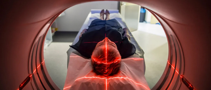
- Imaging Tests: Computed tomography (CT) or magnetic resonance imaging (MRI) scans can reveal potential causes of increased intracranial pressure, such as swelling, bleeding, structural abnormalities, or masses in the brain. Additionally, they can directly visualise brain herniation, confirming the diagnosis & determining the specific type of herniation.
- X-rays of the Skull and Neck: They may be performed to gather additional information.
- Blood Tests: These might be ordered if an abscess or bleeding disorder is suspected as a contributing factor.
Treatment for Brain Herniation
Treatment for brain herniation syndrome requires immediate medical intervention to reduce intracranial pressure and prevent further brain damage. These may include:
- Extraction of Mass Lesions: This may involve surgical removal of haematomas, contusions, tumours, or abscesses that are causing increased pressure within the skull.
- Decompressive Hemicraniectomy: In this process, a portion of the skull is removed to allow more space for the swollen brain.
- Physiological Neuroprotection: This includes sedation and analgesia to calm the patient and reduce metabolic demands on the brain.
- Mechanical Ventilation: It is required to support breathing and control carbon dioxide levels.
- Careful Fluid Management: Preventing further oedema while avoiding dehydration is essential.
- Osmotherapy: It plays a significant role in reducing brain swelling. Drugs (mannitol or hypertonic saline) are administered to draw fluid out of the brain tissue.
- Ventriculostomy: It helps in cerebrospinal fluid drainage to alleviate pressure.
- Other Treatment Options:
- Hyperventilation causes vasoconstriction and lower intracranial pressure
- Therapeutic hypothermia to slow brain metabolism
- Corticosteroids may be used in specific situations, such as tumour-related oedema.
When to See a Doctor
Individuals should go for immediate medical care if they or someone they know exhibits signs of this life-threatening condition, such as:
- Altered consciousness
- Severe headaches
- Dilated pupils
- Drowsiness
- Difficulty concentrating
- Abnormal posturing
- Rigid body movements
- Unusual body positions
Prevention
While it is not always possible to prevent brain herniation, there are several measures individuals can take to reduce their risk. These are:
- People with pre-existing conditions such as brain tumours or blood vessel abnormalities, like aneurysms, are at a higher risk of experiencing brain herniation. Therefore, regular medical check-ups and adherence to prescribed treatments are crucial for these individuals.
- For the general population, the key to prevention lies in avoiding head injuries. This involves:
- Wearing appropriate protective gear
- Refraining from risky behaviours
- Education about the importance of safety measures and the potential consequences of head injuries can encourage individuals to take necessary precautions.
Conclusion
Brain herniation is a serious medical emergency that has a significant impact on patient outcomes. This article has explored the various types, causes, symptoms, and complications of this condition, highlighting the importance of swift diagnosis and treatment. By understanding the risk factors and recognising the warning signs, individuals can take precautionary measures to protect themselves & seek timely medical attention when needed.
FAQs
1. Can you recover from a herniated brain stem?
Recovery from brain stem herniation is challenging and depends on various factors. The severity of the herniation, the underlying cause, and the speed of medical intervention play crucial roles in determining the outcome. Sometimes, prompt treatment can lead to recovery. The brain stem controls vital functions, and damage to this area can have severe consequences. Patients who survive may face long-term neurological issues.
2. Is a herniation reversible?
Brain herniation can be reversible in some cases if treated quickly and effectively. The key lies in addressing the underlying cause and reducing intracranial pressure. For instance, if a brain tumour or haematoma is causing the herniation, surgical removal may alleviate the pressure and reverse the herniation. However, the longer the brain remains herniated, the higher the risk of permanent damage or death.
3. What happens after brain herniation?
After brain herniation occurs, the consequences can be severe and life-threatening. The shifted brain tissue can compress vital structures, leading to a cascade of neurological problems. Patients may experience a rapid decline in consciousness, abnormal breathing patterns, and pupil size and reactivity changes. In severe cases, brain herniation can lead to respiratory failure, cardiac arrest, and, ultimately, brain death if not treated promptly.
4. What is the most common type of brain herniation?
The most common type of brain herniation is subfalcine herniation, also known as midline shift. In this type, brain tissue moves underneath a membrane called the falx cerebri in the middle of the brain, pushing across to the other side. This often occurs due to increased pressure on one side of the brain, typically caused by conditions such as tumours, haematomas, or severe swelling.